Abstract
A Cubital tunnel syndrome caused by hypertrophy of the aberrant muscle, anconeus epitrochlearis in an amateur weight lifter is presented.
Résumé
Un syndrome de tunnel cubital causé par l'hypertrophie du muscle aberrant, anconeus epitrochlearis chez un haltérophile amateur est présenté.
Introduction
We present a case of a cubital tunnel syndrome secondary to the aberrant muscle anconeus epitrochlearis in an amateur weight lifter.
Case report
This 28-year-old right-handed man had an eight-week history of tingling sensation and weakness of the grip power. He denied any trauma to his elbow and his symptoms progressed despite the use of splints and medication for 3 months. He was a businessman and had to do computer works for several hours a day. He attended classes in physical fitness and lifted weight to develop his muscles. Numbness of the 4th and 5th fingers and difficulty in fine motion like touching computer key board became constant recently and weakness of flexor profundus of ulnar 2 digits was present. Examination revealed positive Tinnel sign at the cubital tunnel with radiating pain into the ring and little fingers. However, Tinnel sign was negative at the Guyon's canal. There was no atrophy of the thenar, hypothenar, and interosseous muscles. His symptom was aggravated by elbow flexion beyond 90 degrees. Laboratory test was within normal limit including test for syphilis. Electromyography of the left ulnar nerve showed the prolonged distal latency compared to the opposite (2.46msec: 2.38msec).
The motor amplitude of the ulnar nerve above the elbow was 8.40 compared to 3.37 of the below elbow. Conduction velocity above the elbow was 54.8m/sec compared to 45.9m/sec of the below elbow.
Spontaneous fibrillation of left adductor digiti quinti muscle was observed. In addition, recruitment pattern of left adductor digiti quinti was decreased. Sensory nerve conduction was within normal range. Ultrasound of the cubital tunnel showed no ganglion or mass like lesion, but increased diameter of the ulnar nerve compared to the opposite. He was diagnosed as having an idiopathic cubital tunnel syndrome and planned to surgical release of the cubital tunnel retinaculum.
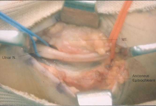
At exploration of the left cubital tunnel, a group of muscle fibers approximately 3-centimeter in width and 3-centimeter in length
(Figure 1)
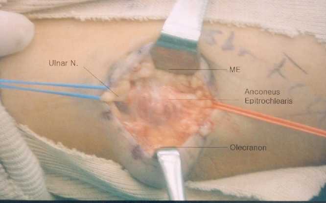
crossed the ulnar nerve from olecranon to medial epicondyle, which was found to be the anconeus epitrochlearis muscle. The ulnar nerve was compressed by the aberrant muscle and a fusiform thickening and induration of the nerve trunk were observed just proximal to the muscle. The muscle was tight in flexion and significantly compressed the ulnar nerve. The ulnar nerve was strained proximal to the muscle bulk at elbow flexion beyond 90. The aberrant muscle was split longitudinally and flexor retinaculum as well. Splitting of the muscle revealed the aberrant muscle was hypertrophied as thick as 5 millimeter in depth and compressed the nerve during elbow flexion. It disclosed narrowed area of ulnar nerve 10mm under the muscle
(Figure 2)
No epineural or perineural neurolysis was performed.
No transposition of the nerve was carried out. Three weeks after surgery, there was gradual recovery and at 6 weeks sensation in the ulnar digits were subjectively and objectively close to normal and his symptoms almost relieved. Electromyography at this time showed improvement in motor nerve conduction velocity and motor distal latency.
Discussion
Various incidence of the anconeus epitrochlearis muscle has been reported in the literature from 34% to 4% in the cadaver study [6,8]. LeDouble [5] reported the muscle present in 32 of 102 cadavers. However, Clemens [2] reported only four cases out of 100 cadavers, one of which was completely developed and other three having only a rudimentary muscle. Although sexes and arms may have nearly equal incidences, the anomalous anconeus epitrochlearis has been reported to be more well developed in men and in the right arm [6]. Hirasawa et al [4] reported similar findings. They reported a weight lifter with bilateral ulnar nerve neuropathy, which was caused by hypertrophy of the muscle. In our study, the patient was also muscular and enjoyed weight lifting.
The boundary for potential ulnar nerve compression begins approximately 10cm proximal to the elbow and end about 5cm distal to the joint. One of them is olecranon groove bounded anteriorly by medial epicondyle, laterally by the olecranon and ulnohumeral ligament, and medially by a fibroaponeurotic covering, which is also called cubital tunnel retinaculum (CTR). This retinaculum has exactly same course as did anconeus epitrochlearis. Thus, O'Driscoll et al [7] considered the CTR as a remnant of the anconeus epitrochlearis muscle and its function is to hold ulnar nerve in position. Compression at this site can be caused by a wide variety of conditions. Aberrant anconeus epitrochlearis is one cause of lesions outside of the groove. In humans, the muscle is probably atavistic and is replaced by a band passing in the same direction as the muscle, called the epitrochleoanconeus ligament.
The treatment for ulnar neuropathy at elbow joint with an associated anconeus epitrochlearis muscle varied from excision of the mass to anterior transposition. Vanderpool et a! [9] and Dahner LE [3] suggested local decompression and excision of the muscle. Although Chalmers [1] recommended anterior transposition of the ulnar nerve, we split the anconeus muscle and local decompression of the ulnar nerve at the cubital tunnel was carried out without anterior transposition. Subluxation has not noted after splitting of the muscle in this study. Masear et al. [6] presented improvement of
electrophysiologic study between 8 and 12 months after operation. We also aware of improvement of electrophysiologic study after surgical release. It is probable that the syndrome can be caused by weight lifting and other overuse condition. If isolated compression by the aberrant muscle, in situ decompression only can achieve satisfactory result.
References
Legends
Figure 1 .